Laboratory Studies
Lamiflo™ Enhances Blood Flow and Tissue Oxygenation
After Stroke and Traumatic Brain Injury.
Neuroprotection for Stroke and Traumatic Brain Injury
Neuroprotection therapies for stroke and traumatic brain injury proven effective in animals all failed clinically. Lamiflo™ offers a new therapeutic approach by improving the physical dynamics of large artery and microvascular blood flow of tissue at risk.*
Lamiflo™ Improves Brain Blood Flow and Tissue Oxygenation After
Acute Ischemic Stroke (pMCAO) and Traumatic Brain Injury (TBI).

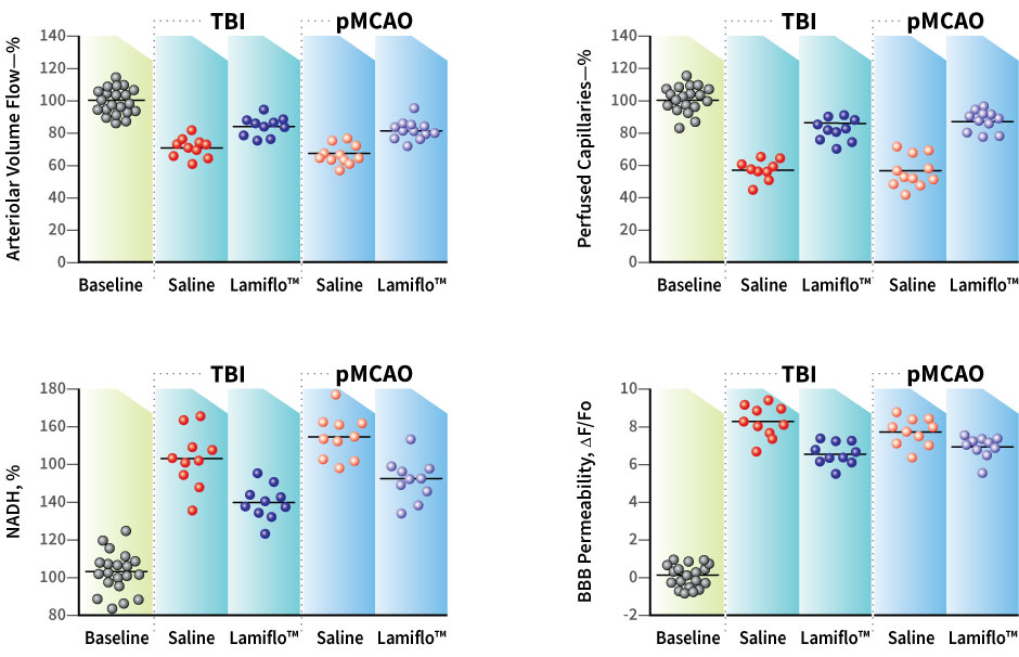
Fig.1) Studies in anesthetized rats, (n=10/group) subjected to permanent middle cerebral artery occlusion (pMCAO) or traumatic brain injury (TBI) studied by microscopy and a fluorescent dye. Variables recorded are percent arteriolar volume flow, perfused capillaries, NADH (lower values=better oxygenation) and blood brain barrier permeability (lower values=lower permeability). Lamiflo™ (2 ug/ml blood) or saline injected i.v. with dye flux via a temporal window insonating the cortical region viewed by 2PLSM at first use. two photon laser scanning microscopy ( 2PLSM). P< 0.05 for all Lamiflo™ and saline group comparisons.
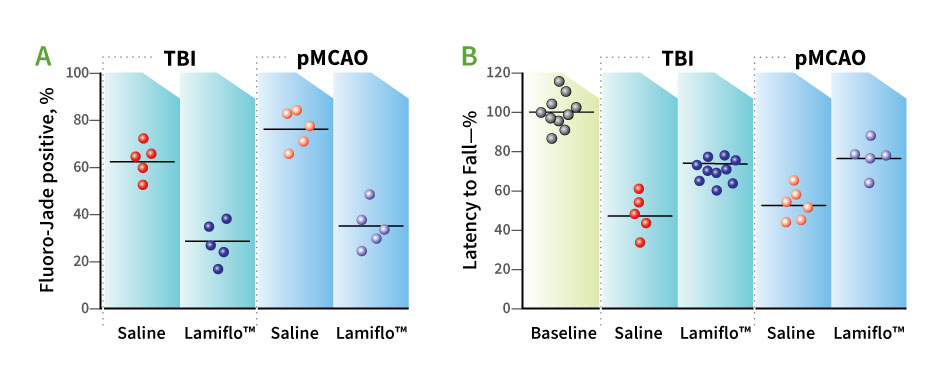
Fig.2) DRP reduces neurodegeneration at 24 hours (A) Fluoro-Jade B stain and (B) improves neurologic outcome by Rotarod 1 week after TBI or pMCAO (N=5 rats per group). Microvascular flow and tissue oxygenation improved by Lamiflo™ (DRP) and tissue oxygenation reduced neurodegeneration by 37.6% (TBI) and 44.2% (pMCAO) compared to saline at 24 hrs by Fluoro-Jade staining, (P<0.05). One week postinsult Lamiflo™(DRP) treated rats performed Rotarod significantly better (P<0.05) (Fig. 2B) than controls.


Fig. 3A) 3-D vasculature reconstruction from Z-stack 2-Photon Laser Scanning Microscopy (2PLSM) images. Capillaries (3-8μm, green) differentiated from larger microvessel (8-20 μm diam, red). B. 2PLSM of rat cortex microvasculature with line scans settings for arteriole blood flow velocity profiles (red stripes). C. Arterial blood flow velocity profiles before [left] and after DRP injection [right, 140 μg/kg 1 hr postTBI] show increased flow velocity near vessel walls (140 μg/kg, 1 h post TBI, Δ,mm/s). D. Imaging volume from where every individual capillary flow was recorded. E. Line scans from the capillary before [up] and after [low] showed increased velocity with DRP. F. Frequency histogram show flow velocity increase after DRP injection. G. Dose effect on capillary flow in male and female rats (for black n=10, for red =3,*P<0.05 Mean Å} SEM).
* Bragin DE, Kameneva MV, Bragina OA, Thomson S, Statom GL, Lara DA, Yang Y, Nemoto EM. Rheological effects of drag-reducing polymers improve cerebral blood flow and oxygenation after traumatic brain injury in rats. J Cereb Blood Flow Metab. 2017 Mar;37(3):762-775.
Bragin DE, Peng Z, Bragina OA, Statom GL, Kameneva MV, Nemoto EM. Improvement of Impaired Cerebral Microcirculation Using Rheological Modulation by Drag-Reducing Polymers. Adv Exp Med Biol 2016;923: 239-244. doi: 10.1007/978-3-319-38810-6_32.PMID: 27526149
